Mast cell tumors (MCT) represent a cancer of mast cells, which are a type of white blood cell involved in allergy and inflammation. Mast cell tumors are the most common skin tumor in dogs. Mast cells contain granules filled with substances that can be released into the bloodstream. In large amounts, these substances can cause systemic (body-wide) complications including stomach ulcers and bleeding and low blood pressure.
Older dogs are most commonly affected. Some predisposed breeds of dogs include Boxers, Boston terriers, Bulldogs, pugs, Labrador retrievers, golden retrievers, Weimaraners, Rhodesian Ridgebacks, cocker spaniels, schnauzers, Staffordshire Terriers, beagles and Shar Peis.
The cause of MCT remains unknown. Because certain breeds of dogs are predisposed to this cancer, genetics are thought to play a role. Additionally, there are known molecular abnormalities associated with MCT. Chronic inflammation may also play a role.
What are the signs and symptoms of mast cell tumors?
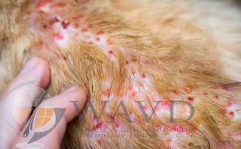
Mast cell tumors can vary in appearance.
These tumors typically present as a lump on or under the skin.
The lump can be hairless or covered by hair, and/or red, ulcerated or swollen.
These lumps are often itchy.
A history of waxing and waning size of the tumor can be reported. This is due to the fact that mast cells contain histamine in their granules and this can be released to cause swelling.
Some MCT can have a more benign-type of behavior where the tumor is present for months to years without change in size or a more aggressive behavior where growth occurs rapidly. If these tumors spread, the typical behavior is first involvement of “local” lymph nodes near the tumor followed by involvement of the liver or spleen.
How is a diagnosis of mast cell tumor made?
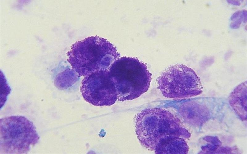
Initial evaluation of a skin mass tends to start with a fine needle aspirate (also called cytology). This is a non-invasive method to extract cells from a mass to obtain a diagnosis. Mast cell tumors have a very specific appearance that should make diagnosis straightforward with cytology.
Following diagnosis of MCT, additional diagnostics performed include:
- Blood work
- Fine needle aspirate of the local lymph node
- Abdominal ultrasound
- Chest radiographs (x-rays)